Image Intensifier
About Image Intensifier
Image Intensifier Services at Nano Hospitals support advanced diagnostic and surgical procedures by providing real-time, high-resolution imaging during medical interventions. This technology enhances X-ray images, allowing doctors to clearly visualize internal structures such as bones, joints, blood vessels, and surgical instruments while performing procedures
Image intensifiers are widely used in orthopaedic surgeries, trauma care, pain management procedures, urology, and various minimally invasive interventions where precision and real-time guidance are essential.
Overview
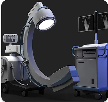
An image intensifier is a medical imaging device, commonly integrated with a C-arm fluoroscopy system, that converts low-intensity X-rays into brighter, visible images. This allows doctors to see moving internal structures in real time during procedures, improving accuracy and safety.
At Nano Hospitals, image intensifier systems are used in operation theatres and procedure rooms to assist specialists in performing precise, minimally invasive treatments.
Workflow
Step 1: Procedure Planning
The doctor determines the need for real-time imaging guidance during a surgical or interventional procedure.
Step 2: Patient Positioning
The patient is positioned on the procedure table, and the C-arm machine is aligned to capture the required anatomical area.
Step 3: Real-Time Imaging
Low-dose X-rays pass through the body, and the image intensifier converts them into clear live images displayed on monitors.
Step 4: Guided Intervention
Doctors use the live images to guide instruments, implants, injections, or catheters with high precision.
Step 5: Post-Procedure Review
Images are reviewed to confirm correct placement or treatment outcome before completing the procedure.
Benefits & Value
For Patients
More accurate and minimally invasive procedures
Smaller incisions and faster recovery times
Reduced risk of complications due to precise guidance
For Doctors
Real-time visualization of internal anatomy
Improved accuracy in implant placement and interventions
Better clinical outcomes in complex procedures
For the Community
Access to advanced image-guided treatments locally
Reduced need for large open surgeries
Improved trauma and emergency surgical car
Risks & Challenges
- • Exposure to low levels of radiation (kept within safe limits)
- • Need for strict radiation protection measures
- • Technical precision required for optimal image quality
- • Limited use in certain patients depending on condition
Frequently Asked Questions