Ultrasound
About Ultrasound
Ultrasound Services at Nano Hospitals provide safe, non-invasive imaging to visualize internal organs, blood flow, and developing fetuses using high-frequency sound waves. This radiation-free technique supports early diagnosis, pregnancy monitoring, and guidance for various medical procedures.
Ultrasound plays a key role in evaluating abdominal organs, pelvic structures, thyroid, breast, soft tissues, and vascular conditions.
Overview
Ultrasound imaging uses high-frequency sound waves that bounce off internal structures to create real-time images on a monitor. It is widely used for examining the liver, kidneys, gallbladder, uterus, ovaries, prostate, thyroid, and soft tissues. Doppler ultrasound additionally assesses blood flow in arteries and veins.
At Nano Hospitals, ultrasound examinations are performed using modern high-resolution machines for accurate and timely diagnosis.
Workflow
Step 1: Doctor’s Referral
A physician recommends ultrasound to investigate pain, swelling, pregnancy, organ function, or suspected abnormalities.
Step 2: Preparation
Depending on the scan type, patients may need to fast or have a full bladder for better image clarity.
Step 3: Gel Application
A water-based gel is applied to the skin over the area being examined to help transmit sound waves.
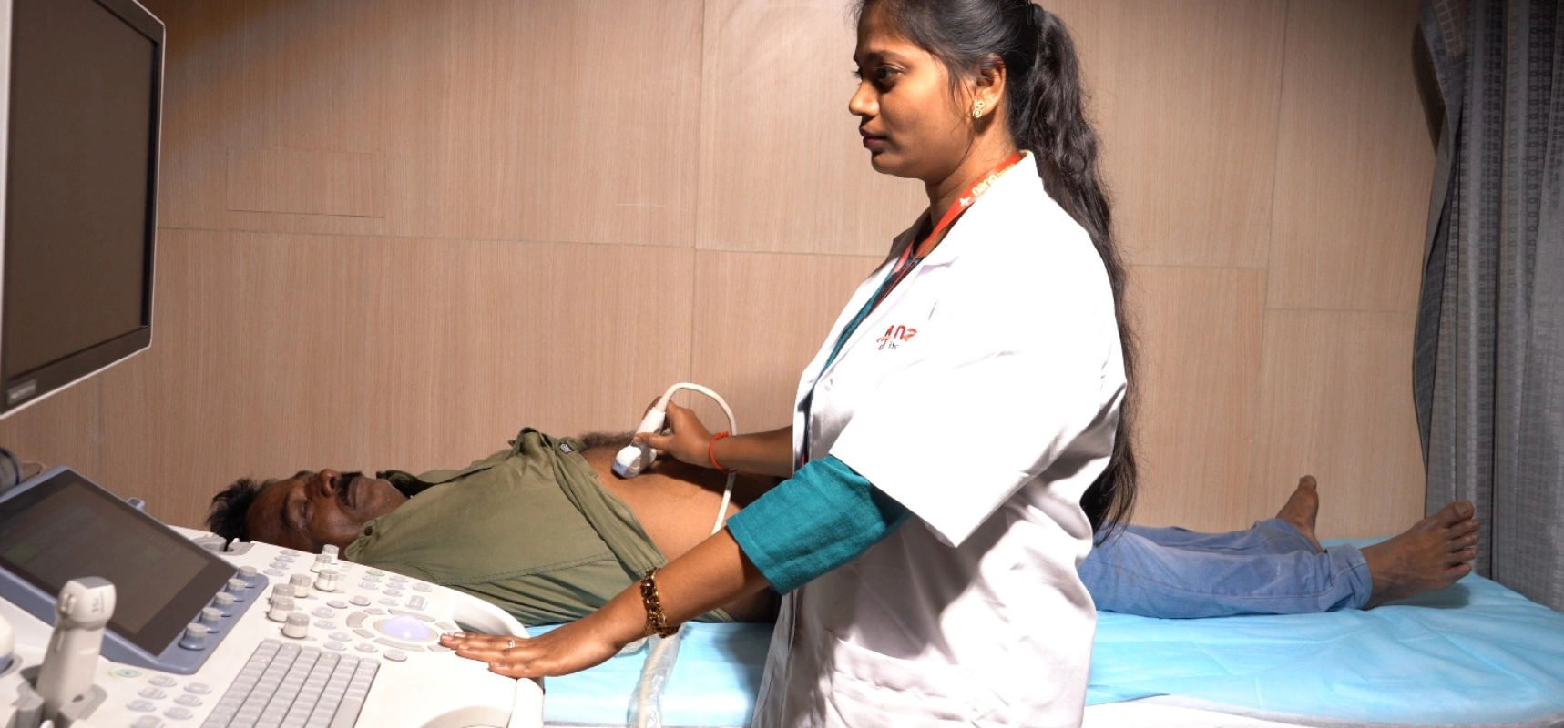
Step 4: Image Acquisition
The radiologist or sonographer moves a handheld probe over the skin to capture real-time images of internal organs.
Step 5: Interpretation and Report
Images are analysed by a radiologist, and a diagnostic report is prepared for the treating doctor.
Benefits & Value
For Patients
Safe imaging without radiation exposure
Painless and non-invasive procedure
Early detection of organ and soft tissue conditions
For Doctors
Real-time visualization of internal structures
Guidance for procedures like biopsies and fluid drainage
Reliable monitoring of pregnancy and fetal development
For the Community
Accessible diagnostic imaging for various medical needs
Early disease detection and preventive care support
Improved maternal and fetal health monitoring
Risks & Challenges
- • Generally very safe with no known harmful radiation
- • Image quality may be limited in obese patients or excessive bowel gas
- • Some scans require preparation such as fasting or a full bladder
Frequently Asked Questions