




2D Echocardiography
Nano Hospitals offers 2D Echocardiography in Bangalore, a non-invasive, painless ultrasound test that evaluates heart chamber size, valve function and pumping strength in 20–40 minutes. Studies are performed and interpreted by cardiologists, with results integrated into cardiac care and emergency/ICU pathways at both Bannerghatta Road and Uttarahalli branches.
About 2D Echocardiography
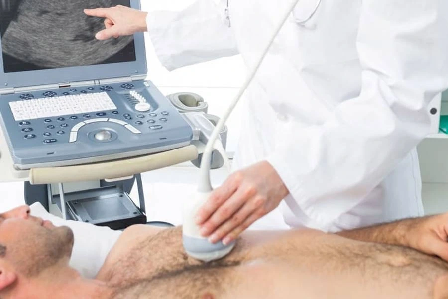
2D Echocardiography (2D Echo) is a non-invasive ultrasound examination of the heart that uses sound waves to create real-time images of cardiac structure and function
It helps doctors assess how the heart chambers, valves, and muscles are working-making it a vital test for diagnosing and monitoring a wide range of heart conditions.
Overview
2D Echo at Nano Hospitals follows a simple, patient-friendly clinical process:
- Step 1: Clinical Indication The treating doctor recommends a 2D Echo based on symptoms such as chest pain, breathlessness, palpitations, or abnormal ECG findings.
- Step 2: Image Acquisition An ultrasound probe is placed on the chest to capture real-time images of the heart from different angles.
- Step 3: Functional Assessment Heart size, pumping strength, valve function, and blood flow patterns are evaluated.
- Step 4: Interpretation The cardiologist analyses the images to identify abnormalities or confirm normal heart function.
- Step 5: Care Planning Results are integrated into the patient’s treatment or follow-up plan.
Workflow
- Step 1: Positioning Patient lies on side.
- Step 2: Probe Placement Probe placed on chest.
- Step 3: Imaging Heart structures viewed.
- Step 4: Measurements Function assessed.
- Step 5: Report Findings recorded.
Benefits & Value
For Patients
Early detection of heart disease
Non-invasive, painless evaluation
Better treatment planning and monitoring
For Families & Caregivers
Clear understanding of heart health
Reduced anxiety through timely diagnosis
Confidence in evidence-based cardiac care
For the Community
Improved early identification of cardiac conditions
Support for preventive and long-term heart care
Reduced complications through timely intervention
Risks & Challenges
2D Echo is a very safe procedure with minimal limitations:
Image quality may vary depending on the patient
Patients may need to change position during the scan
Risk mitigation at Nano Hospitals includes:
Multiple views are taken for better assessment
Experienced cardiologists review the results
Findings are correlated with other tests when required
Frequently Asked Questions
What is a 2D Echocardiography test? +
A 2D Echo is a non-invasive test that uses ultrasound waves to create moving images of the heart. It helps doctors assess the heart’s structure and function.
Why is a 2D Echo recommended? +
It may be recommended to evaluate symptoms such as chest discomfort, breathlessness, irregular heartbeat, or to check for heart-related conditions.
Is 2D Echocardiography painful? +
No. A 2D Echo is generally painless and does not involve needles, radiation, or surgery.
How long does a 2D Echo take? +
The test usually takes around 20–40 minutes, depending on the type of examination required.
Do I need to fast before a 2D Echo? +
Usually, fasting is not required for a standard 2D Echo. You can generally eat and drink normally unless your doctor gives you different instructions.
Is any preparation required before the test? +
Usually, no special preparation is needed. You may be asked to remove clothing from the upper body so the ultrasound probe can be placed on the chest.
Is 2D Echo safe? +
Yes. A standard 2D Echo uses ultrasound waves and does not expose you to ionizing radiation. It is generally considered a safe diagnostic test.
What can a 2D Echo detect? +
It can help assess heart chambers, valves, pumping function, blood flow, and certain structural abnormalities of the heart.
Can I resume normal activities after a 2D Echo? +
Yes. Since a standard 2D Echo is non-invasive, most people can return to their normal activities immediately after the test.
When will I receive my 2D Echo report? +
The report timing can vary depending on the hospital and examination. Your healthcare team at Nano Hospitals can provide information about when your report will be available.
How can I book a 2D Echocardiography appointment at Nano Hospitals? +
Yes, Ankle-Brachial Index (ABI) testing is available at Nano Hospitals Bangalore by appointment. Patients can book an ABI test by calling the hospital reception desk or using the online appointment booking option at abi. Please call Nano Hospitals before visiting to confirm appointment availability and timings.