Imaging for Foot (X-ray, MRI)
About Imaging for Foot (X-ray, MRI)
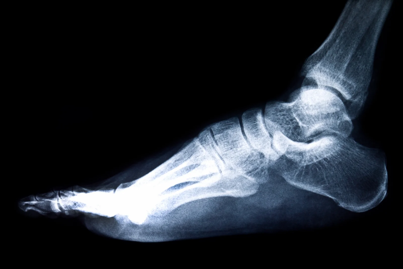
Imaging facilities such as X-ray and MRI help evaluate bone, soft tissue, an...
Imaging facilities such as X-ray and MRI help evaluate bone, soft tissue, and infection-related complications in diabetic feet.
Overview
Imaging is important for detecting fractures, osteomyelitis, Charcot foot changes, and deep tissue infections.
Understanding The Procedure
Symptoms Imaging for Foot (X-ray, MRI)
- ⦾ Symptoms or clinical findings that require Imaging for Foot (X-ray, MRI) for clearer diagnosis
- ⦾ Unresolved or unexplained complaints after initial clinical evaluation
- ⦾ Need to confirm disease extent, severity, or organ involvement
- ⦾ Monitoring of a known condition where imaging or testing guides next steps
- ⦾ Pre-treatment planning where accurate assessment is required
Recovery & Outlook
Early imaging evaluation improves diabetic foot treatment outcomes and helps prevent severe complications.
Risks
- ⦾ MRI restrictions in some patients
- ⦾ Radiation exposure with X-rays
- ⦾ Cost of advanced imaging
Post-Operative Care
- ⦾ Follow specialist recommendations
- ⦾ Continue wound care and medications
- ⦾ Attend follow-up imaging if required
Prefer WhatsApp for Imaging for Foot (X-ray, MRI) enquiries? We respond on chat during working hours.