




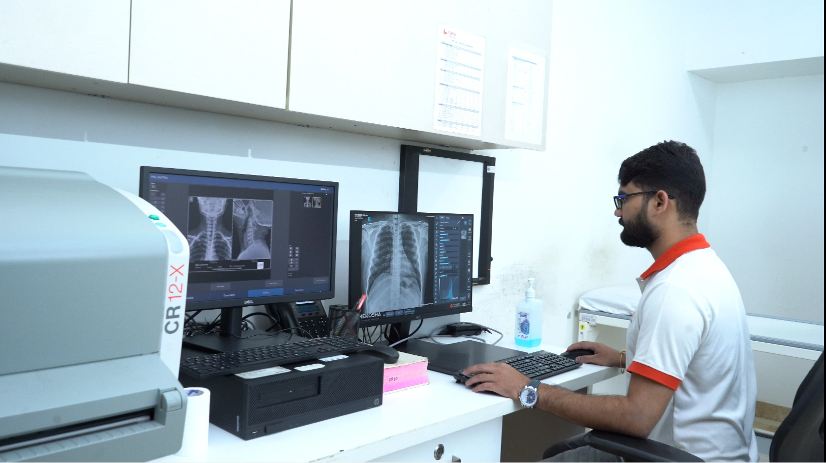
Digital X-Ray
Nano Hospitals provides digital X-ray services in Bangalore to create images of internal body structures for medical evaluation. These images help doctors assess and diagnose medical conditions.
About Digital X-Ray
Digital X-Ray is a fast imaging test used to evaluate bones, chest, joints, and injuries. It produces instant images with minimal radiation exposure.
It helps diagnose fractures, infections, lung diseases, and structural abnormalities quickly.
Overview
The digital system provides high-quality images immediately on computer screens for rapid diagnosis.
Doctors can review and share reports instantly for treatment decisions.
Workflow
- Step 1: Preparation Patient positioned and metal objects removed.
- Step 2: Image Capture X-ray beam passes through body part.
- Step 3: Digital Processing Images appear instantly on monitor.
- Step 4: Quality Check Technician verifies clarity.
- Step 5: Reporting Radiologist reviews and reports.
Benefits & Value
For Patients
Quick procedure
Minimal radiation
Immediate diagnosis
For Caregivers & Families
Faster treatment planning
Less waiting time
Clear explanation
For the Community
Early fracture detection
Faster emergency care
Accessible imaging
Risks & Challenges
Minor radiation exposure
Movement blur
Positioning difficulty
Pediatric cooperation
Frequently Asked Questions
What is a Digital X-Ray at Nano Hospitals? +
Digital X-Ray at Nano Hospitals is an advanced imaging service that uses X-rays to produce clear digital images of bones and other internal structures of the body.
What is Digital X-Ray used for at Nano Hospitals? +
Digital X-Ray at Nano Hospitals helps doctors evaluate fractures, bone injuries, chest conditions, joint problems, and other abnormalities.
Which body parts can be examined using Digital X-Ray at Nano Hospitals? +
Digital X-Ray at Nano Hospitals can be used to examine the chest, spine, arms, legs, joints, and other areas as recommended by the doctor.
How does Digital X-Ray work at Nano Hospitals? +
At Nano Hospitals, X-rays pass through the body and are captured by a digital detector to produce images that can be reviewed by the radiology team.
How long does a Digital X-Ray take at Nano Hospitals? +
A Digital X-Ray at Nano Hospitals is generally a quick procedure, with the actual imaging usually taking only a few minutes depending on the area being examined.
Is Digital X-Ray at Nano Hospitals painful? +
No. Digital X-Ray at Nano Hospitals is a non-invasive procedure and does not cause pain. You may simply need to remain in a specific position while the image is taken.
Do I need any preparation for a Digital X-Ray at Nano Hospitals? +
Most Digital X-Rays at Nano Hospitals require little or no special preparation. You may be asked to remove jewellery or metallic objects before the examination.
Is Digital X-Ray at Nano Hospitals safe? +
Yes. Digital X-Ray at Nano Hospitals uses controlled radiation exposure, with appropriate safety measures followed during the examination.
How are Digital X-Ray results provided at Nano Hospitals? +
The images are produced digitally and reviewed by the radiology team at Nano Hospitals. The reporting time may vary depending on the type of examination.
Can we book an appointment for Digital X-Ray at Nano Hospitals? +
Yes. Digital X-Ray services at Nano Hospitals Bangalore can be scheduled by appointment through digital-x-ray, by calling the hospital, or by visiting the hospital reception desk. Digital X-Ray services are available at both Bannerghatta Road and Uttarahalli branches.